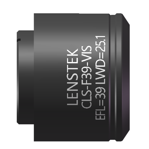
| CLS-F40-VIS | |
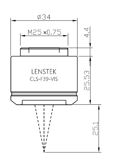
| Wavelength | 400nm-700nm |
| Effective Focal Length | 39mm |
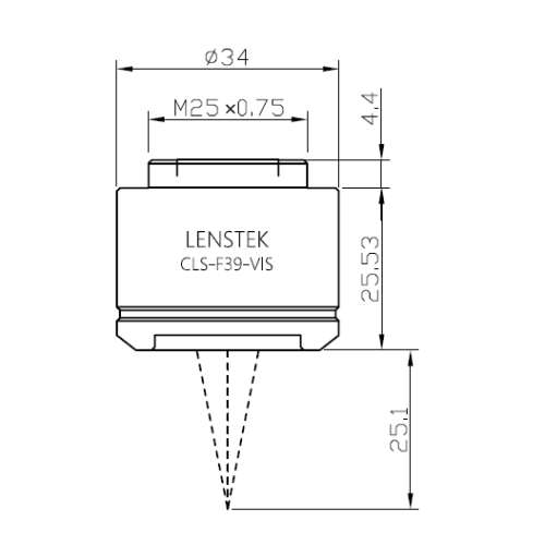
| Working Distance | 25.1mm |
| Field of View | 10.3mmx10.3mm |
| Diffraction Limited | 9mmx9mm |
| Mounting Threads | M25X0.75 |
| Entrance Pupil Position | 25mm |
| Entrance Pupil Diameter | 5mm |
The CLS-F40-VIS is a visible light scanning lens with an operating wavelength range of 400–700 nm and an effective focal length of just 39 mm. Thanks to its short focal length design, this lens achieves a higher numerical aperture and a smaller diffraction-limited spot size within a compact optical system, significantly enhancing lateral resolution and image detail rendition. It is specifically optimized for OCT (Optical Coherence Tomography) imaging systems while being widely applicable to confocal microscopy, light sheet microscopy, high-content screening systems, and other laser scanning microscopy applications. In OCT systems, the short 39 mm focal length enables a finer focal line or smaller spot size at the sample, directly improving both axial and lateral resolution. This results in clearer tomographic images for preclinical and clinical applications such as retinal imaging, dermatological examination, and cardiovascular endoscopy.
The CLS-F40-VIS delivers diffraction-limited performance across the entire visible band (400–700 nm). This means that wavefront aberrations are corrected to approach the theoretical physical limit, and the energy distribution of the focused spot is strictly governed by diffraction without additional geometric blurring. As a result, each scanned point achieves the minimum possible spot size dictated by the system, yielding sharp, high-contrast microscopic images. This performance is particularly critical for fluorescence imaging requiring precise photon localization and for detecting weak signals in scattering media.
The lens features an entrance pupil diameter of 5 mm and supports a maximum field of view of 10.3 mm × 10.3 mm, making it suitable for large-area tissue imaging or high-throughput multi-well plate scanning. The key enabling factor is that when used with our standard tube lens (such as the TTL200 series), the system forms an object-space telecentric optical configuration. In a telecentric path, the chief rays are parallel to the optical axis as they enter the sample plane. Consequently, regardless of whether the scanning beam is at the center or the edge of the field of view, the magnification and spot shape remain constant. This fundamentally eliminates vignetting and illumination roll-off at the field edge, ensuring uniform brightness and shadow-free imaging across the entire 10.3 mm × 10.3 mm field — an essential feature for quantitative analyses such as fluorescence intensity statistics and OCT signal attenuation correction.
Additionally, the CLS-F40-VIS is equipped with a standard M25×0.75 external thread for easy integration into most commercial or custom scan heads. The broadband anti-reflection coating for the visible range achieves an average reflectance of <0.5% from 400 to 700 nm, effectively suppressing stray light and improving image signal-to-noise ratio. Whether you are building a compact OCT system or upgrading an existing laser scanning microscope, the CLS-F40-VIS delivers clear, large-field, and reproducible imaging results through its short focal length, diffraction-limited image quality, and true telecentric uniformity.
Applications:
Optical Coherence Tomography (OCT) imaging systems
Confocal microscopy
Light sheet microscopy
High-content screening systems
General laser scanning microscopy

| LAYOUT | ZEMAX FILE | 3D STEP |
CLS-F40-VIS
- Product Code: CLS-F40-VIS
- Availability: In Stock
-
Â¥7,397.00
- Ex Tax: ¥7,397.00